The application of physics principles, techniques, and strategies in clinical exercise and investigate has revolutionized the overall health-related science field to increase human wellbeing and all round wellbeing.

Impression Credit history: Nuttawut Yeenang/Shutterstock.com
What is Health care Physics?
Professional medical physics is a department of utilized physics that utilizes bodily sciences to stop, diagnose, and deal with human illnesses. Clinical physics can be classified into several sub-groups: medical imaging physics, radiation oncology physics, non-ionizing health care radiation physics, nuclear drugs physics, health-related overall health physics, and physiological measurements.
Health-related physics largely focuses on ionizing radiation measurement, magnetic resonance imaging, and making use of physics-centered systems (lasers and ultrasound) in medicine.
The term “healthcare physics” was initially launched by Félix Vicq d’Azir, a French physician, anatomist, and the typical secretary of the Royal Society of Medicine, in Paris in 1778. In 1814, the most correct definition of clinical physics was launched in the revised version of Nysten’s medical dictionary. In this version, health care physics was described as “physics used to the understanding of the human human body, to its preservation and to the overcome of its diseases.”
What is Health care Physics?
Vital Roles of Medical Physicists?
Health-related physicists are health care industry experts who have specialized schooling in applying physics ideas and systems in medication. They largely operate in medical setups or in tutorial and study establishments. The vital roles and duties of health-related physicists incorporate the application of healthcare physics methods for the diagnosis and treatment of human health conditions and the security of professional medical staff and individuals from ionizing and non-ionizing radiation dangers.
Clinical physicists specialized in radiation remedy are mainly associated in giving radiation therapies for cancer patients in collaboration with oncologists and other therapists. The remedies mostly involve brachytherapy, whereby a radiation source is put inside the human body, or exterior beam radiation therapy, whereby linear accelerator-generated radiation is thoroughly shipped to affected tissues.
Health-related physicists specializing in medical imaging are engaged in developing and keeping various imaging tactics, such as x-ray, computed tomography scan (CT-scan), and magnetic resonance imaging.
Clinical physicists specialized in nuclear physics largely conduct practical imaging of people making use of positron emission tomography (PET), gamma digital camera, and organic substances labeled with radioactive markers (radiopharmaceuticals).
X-Ray and CT Scan
In X-rays, alerts produced from a slender X-ray beam transverse the influenced space of fascination to make planer photos. Similarly, cross-sectional X-ray photos obtained upon recurring scanning are digitally stacked to create substantial-resolution, a few-dimensional, or four-dimensional computed tomography (CT) photos to evaluate dynamic processes.
Moreover supplying quantitative and reproducible anatomical photos, CT can deliver superior-quality useful information and facts by means of dynamic perfusion scanning. In the course of the perfusion treatment, a contrasting agent is administered, and repeated imaging of the afflicted area is done at an interval of 3 – 5 seconds for 30 seconds. These pictures are subsequently stacked to type four-dimensional illustrations or photos. This strategy is very practical in analyzing hemodynamic parameters, such as blood circulation and blood volume.
Magnetic Resonance Imaging
Magnetic resonance imaging (MRI) is a impressive non-invasive medical imaging procedure that makes use of a strong, static magnetic discipline, magnetic gradients, and computer system-induced radio waves to deliver high-top quality 3-dimensional visuals of tissues and organs. The magnetic discipline utilized to the entire body realigns the body’s photons with that subject. Subsequently, radio waves promote photons, and MRI sensors are utilised to detect power (sign) launched from photons.
In quantitative MRI, contrast distinctions between two tissues are maximized on a solitary picture by utilizing the peace time variances of two tissues. The photographs are weighted centered on the properties of one particular tissue. The modalities frequently applied for quantitative MRI include things like arterial spin labeling for cerebral blood move measurement and diffusion tensor imaging for microstructural investigation.
Ultrasound
Ultrasound is a superior-frequency audio wave that generates non-invasive photographs of distinct tissues and organs. The variance in mechanical properties at the interface of different organs/tissues brings about ultrasound reflection. These reflections are measured to make ultrasound pictures.
The major pros of ultrasound about other clinical imaging procedures (CT and MRI) are value-performance and genuine-time imaging at the bedside. Distinction boosting agents, these types of as microbubbles, are applied in ultrasound for practical imaging. Aside from disorder prognosis, ultrasound is utilized for therapeutic functions. For instance, higher-intensity focused ultrasound eliminates influenced tissues inside of the entire body without having harmful bordering healthy tissues. In addition, ultrasound is utilized for specific drug shipping and delivery.

Image Credit rating: Output Perig/Shutterstock.com
Nuclear Drugs
In nuclear drugs, radioactive probes are applied to notice physiological processes. The probes are also used for targeted delivery of therapeutic doses. A very tiny sum of radioactive probe is administered to the body for the duration of the course of action. The probe is subsequently absorbed by the organ/tissue less than investigation. The radiation emitted from the probe thanks to decay is detected by a gamma digicam, which generates digital signals for examining the practical point out of the organ.
A gamma digital camera generates two-dimensional photographs when it remains stationary. In one-photon emission computed tomography, the digital camera is rotated to generate axial slices of the target organ. These slices can be applied in PET scans to make a few-dimensional photographs.
Radiotherapy
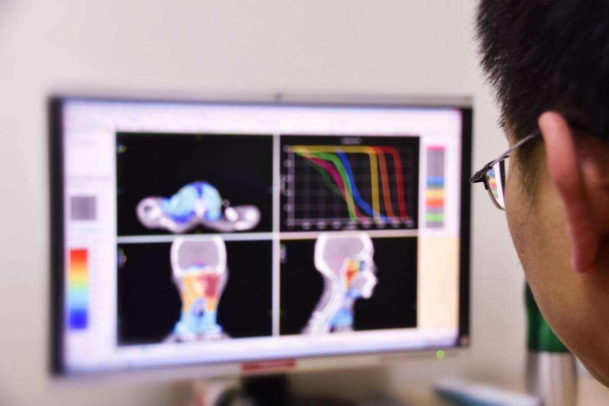
Radiation treatment entails the delivery of ionizing radiation within the overall body to demolish and get rid of most cancers cells. For deep-seated tumors, significant-electrical power photons are applied. For superficial tumors, higher-strength electrons are made use of. In addition, billed particles, together with protons, are made use of in radiotherapy.
Throughout the full cure treatment, professional medical imaging is performed to make certain risk-free and focused delivery of the radiation and to assess radiation-induced improvements in the anatomy.
References
Additional Examining
